Custom AI development
Vision Transformers, UNet, Segformer, DeeplabV3 and etc. for segmentation and biomarker discovery. Deliverables: .pt checkpoints, inference benchmarks and validation reports.
DIGITAL PATHOLOGY × SPATIAL BIOLOGY
Digital pathology, spatial biology and custom AI — connected in one reproducible research workflow. Turn whole-slide images into biologically meaningful, publication-ready results.
Discuss your research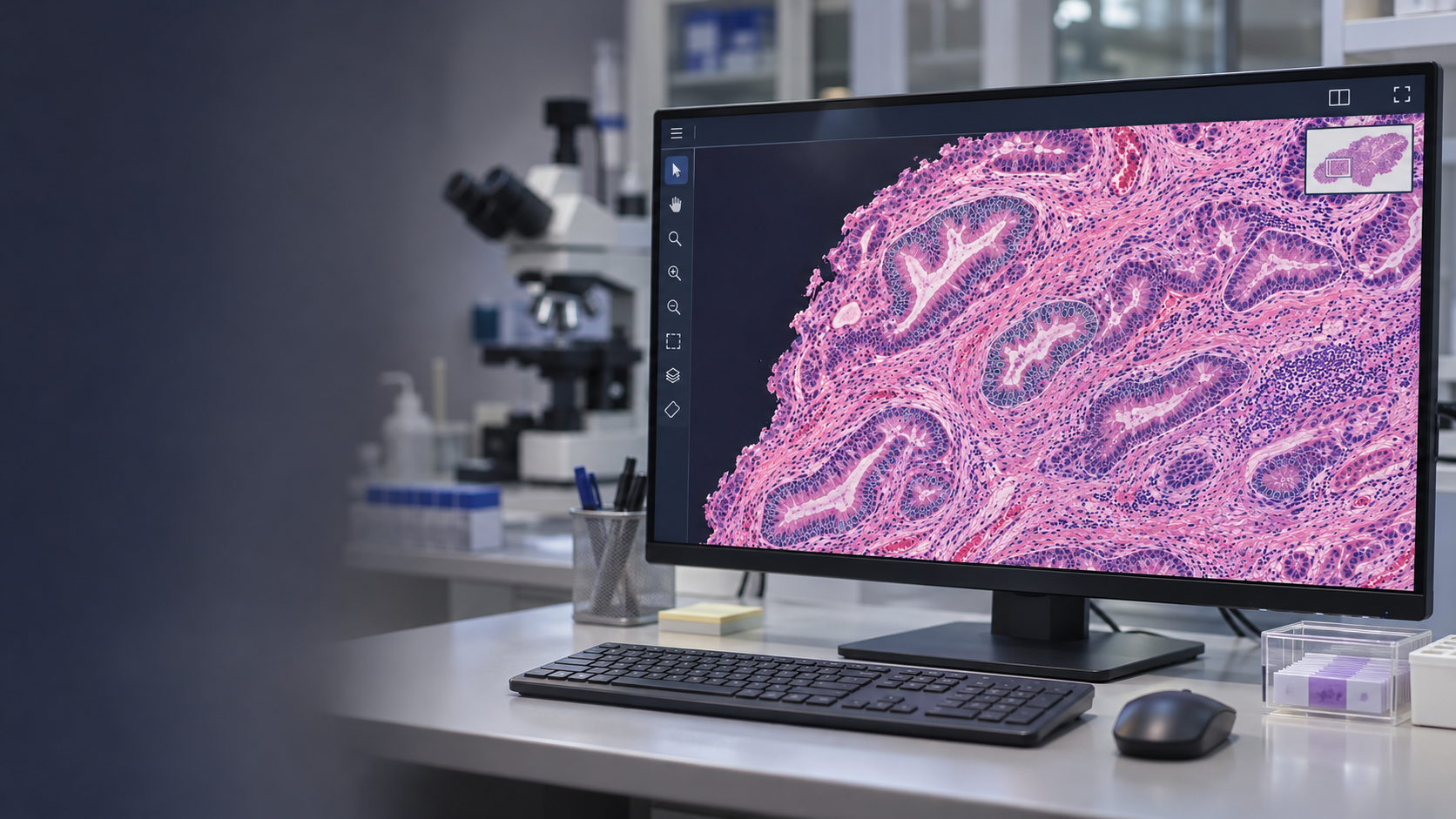
01 / SCIENCE, CONNECTED
We bring pathology, computer vision, deep learning, spatial biology, and biostatistics into a unified research platform.
Work with us as a full scientific partner or bring our expertise into a specific stage of your study. Every workflow is tailored to your scientific objectives — with reproducibility, rigor, and expert pathology validation at its core.
02 / OUR EXPERTISE
Custom algorithms. Quantitative evidence.
Biologically meaningful interpretation.
Vision Transformers, UNet, Segformer, DeeplabV3 and etc. for segmentation and biomarker discovery. Deliverables: .pt checkpoints, inference benchmarks and validation reports.
Measure nuclear pleomorphism, stroma-to-tumor ratio, digital H-scores. Export objective morphometry tables with expert pathology review.
Delaunay triangulation, nearest-neighbor distances and cellular density gradients. Deliverables: single-cell coordinates, interaction heatmaps and GeoJSON clusters.
Affine and elastic serial-section alignment with ROI transfer across stains. Deliverables: aligned image stacks and synchronized multi-stain review.
Integrate CD3, CD8, CD20, CD68, FOXP3 and other markers. Characterize immune infiltration, invasive margins and tertiary lymphoid structures (TLS).
QuPath, OpenSlide, Python and PyTorch workflows, containerized with Docker / Singularity for on-premise HPC or private cloud. Code, recipes and execution provenance included.
OUR PROJECTS / RESEARCH SOFTWARE
We’ve developed tools to support researchers’ day-to-day work in tissue segmentation, slide alignment and marker comparison.
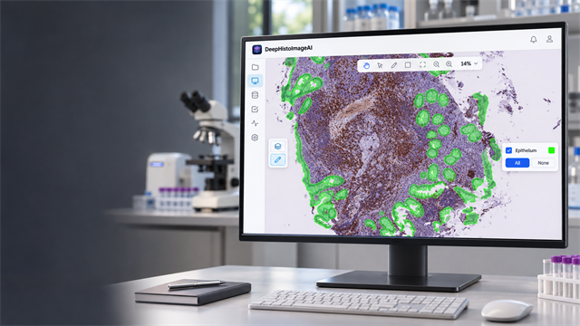
01 / SEGMENTATION · WINDOWS
Annotate tissue, train a segmentation model on your own data and review prediction masks — without writing ML code. Original slides stay local; export results to QuPath via GeoJSON.
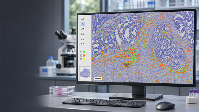
02 / ALIGNMENT · DESKTOP
Align adjacent tissue sections stained for different markers. Transfer annotations and cell contours, compare staining intensity and export GeoJSON to QuPath.
03 / THE RESEARCH WORKFLOW
A standardized computational foundation connects your data, methods, and results — from pilot studies to multicenter cohorts with thousands of slides.
Frame the protocol and power calculation plan. Ingest whole-slide images in SVS, NDPI, CZI and other formats.
Detect blur, folds, bubbles and pen marks. Generate tissue masks; normalize stains with Macenko, Vahadane or Reinhard.
Align serial sections, transfer multi-marker ROIs and annotate the tissue architecture relevant to your hypothesis.
Segment nuclei and cells, classify phenotypes, quantify spatial distances and identify microenvironment clusters.
Compare patient groups and spatial biomarkers to see how they relate to survival.
Expert pathology review, Methods and Results drafts, SHA-256 provenance and publication-ready vector / 300+ DPI TIFF figures.
Quality control at every stage. Traceable from input to figure.
RESEARCH / PUBLICATIONS
Research in digital pathology, tissue analysis and spatial biology.
Author names · Journal · Year
Author names · Journal · Year
0102
04 / WORK WITH US
A connected scientific partnership from the first research question to the final manuscript.
Study design · Image analysis · AI & spatial modeling · Biostatistics · Publication support
Specialist support that integrates into your existing study, team, or computational infrastructure.
Custom AI · Image registration · Spatial analysis · Software development · Expert validation
Single-cell CSV / Parquet tables, GeoJSON annotations, QuPath .qpdata, aligned masks, model checkpoints and 300+ DPI figures — with transparent Methods and reproducible code.
05 / REPRODUCIBILITY & DATA STEWARDSHIP
Institutional anonymization and NDA-led data intake. Agree storage, access and retention with your research team.
SHA-256 input hashes and versioned pipeline parameters connect each analysis to its source data.
Containerized execution for on-premise HPC or private cloud. Define encryption and institutional controls during scoping.
06 / START A RESEARCH PROJECT
Tell us about your tissue, hypothesis and timeline. We’ll review feasibility and suggest a computational approach.